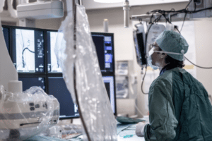
A combined team of researchers from the University of California and Oregon Health and Science University has developed a microelectrode array to precisely monitor electrophysiology during spinal surgery. They have published their work in the journal Science Translational Medicine.
Surgery on the spine is a complicated procedure. In addition to correcting the problem, doctors should do it without harming the patient’s neurons, which could result in disabilities. Tumor excision is also an area that needs special attention, but even taking biopsies is dangerous. Nowadays, intraoperative neuromonitoring tools are used by spinal surgeons to track nervous system electrical impulses.
Electrodes are applied to the scalp and back muscles using such gadgets. However, they don’t offer the accurate data that surgeons want. They frequently only help the surgeon recognize the injury that has previously happened. In this new endeavor, the scientists have made an effort to enhance the technology by developing a tool that enables more accurate electrophysiological tracking throughout the operation.
The new gadget comprises an arrangement of electrodes that the neurosurgeon implants proximally to the main surgical site on the spinal column. The electrodes monitor the electrophysical activity of the nerve fibers in the spine and communicate that information to a computer, which transforms it into a submillimeter-accurate 2D map that the neurosurgeon can view in real-time while performing surgery. The scientists used volunteers to test their novel equipment during six real surgeries. They discovered that their gadget beat existing ones in helping the neurosurgeon during spinal procedures and offered more responsiveness than they did.
Before the gadget can be filed for authorization, more test is needed, but the scientists are convinced their approach will give spinal surgeons more reliable data and hence minimize brain injuries in future patients. Additionally, they believe that patients with injured spinal columns might use their gadget to track the development of their recuperation.
Access Planning, and skills of learning
The very first crucial stage in spinal endoscopy is creating an access point. The initial guidewire is inserted for the interlaminar method using a spinal needle at the intersection of the operative level’s facet joint complex and the lower side of the rostral lamina. To address the lateral spinal canal through a transforaminal technique, a spinal needle is used to push the protective into the triangular safe area between the outgoing and traversing nerve roots. The ideal angles for accessible paths range for L3-4 is from 10° to 40° in the axial plane, for L4-5 is 55° to 65° in the sagittal plane, and for L5-S1 is 25° to 50° in the coronal plane.
Ergonometric Systems and training
In order to ease decompression movements through the tiny internal operating path of the spinal endoscope, endoscopic tools are developed around the endoscopic platform. The majority of modern foraminoscopes made for transforaminal decompression contain an inner working canal of around 4.3 mm to fit common neurosurgical tools, most of which have been adapted to fit through lengthy endoscopes.
For translaminar endoscopic methods, like those used during interlaminar surgery to accelerate the decompression, large inner diameter endoscopes of 7.1 mm are used. To enable the neurosurgeon to use both hands skilfully, these tools must be ergonomically designed. The endoscope is handled in the left hand of a right-handed neurosurgeon with the forefinger touching the operating channel.
Challenging Clinical Scenarios
A beginning spine surgeon should take on a number of difficult contingencies until they are proficient enough to do so. Large paracentral, central, foraminal, up- or down-migrated, or far-lateral disc herniations fall under this category. Additionally, a novice should avoid treating large facet cysts. Dural and nerve root sleeve damage should be anticipated, as are dural adhesions, which are frequent.
Herniations caused by calcified discs may also be challenging to treat and frequently call for the usage of power tools. It is normal to anticipate many calcifications. Doctors must decide for themselves if a particular situation is outside of their scope of practice. These more serious herniations or extreme stenosis situations may be less difficult to manage with training and experience.


